Tuesday, May 19, 2009

Night vision range

Spectral range

Night-useful spectral range techniques make the viewer sensitive to types of light that would be invisible to a human observer. Human vision is confined to a small portion of the electromagnetic spectrum called visible light. Enhanced spectral range allows the viewer to take advantage of non-visible sources of electromagnetic radiation (such as near-infrared or ultraviolet radiation). Some animals can see well into the infrared and/or ultraviolet compared to humans.

Intensity range

Sufficient intensity range is simply the ability to see with very small quantities of light. Although the human visual system can, in theory, detect single photons under ideal conditions, the neurological noise filters limit sensitivity to a few tens of photons, even in ideal conditions.[2]

Many animals have better night vision than humans do, the result of one or more differences in the morphology and anatomy of their eyes. These include having a larger eyeball, a larger lens, a larger optical aperture (the pupils may expand to the physical limit of the eyelids), more rods than cones (or rods exclusively) in theretina, a tapetum lucidum, and improved neurological filtering.

Enhanced intensity range is achieved via technological means through the use of an image intensifier, gain multiplication CCD, or other very low-noise and high-sensitivity array of photodetectors.

Monday, May 18, 2009

Night glasses


Binoculars (night vision goggles on flight helmet) Note: the green color of the objective lenses is the reflection of the Light Interference Filters, not a glow.

Night glasses

Night glasses are telescopes or binoculars with a large diameter objective. Large lenses can gather and concentrate light, thus intensifying light with purely optical means and enabling the user to see better in the dark than with naked eye alone. Often night glasses also have a fairly large exit pupil of 7 mm or more to let all gathered light into the user's eye. However, many people can't take advantage of this because of the limited dilation of the human pupil. To overcome this, soldiers were sometimes issuedatropine eye drops to dilate pupils. Before the introduction of image intensifiers, night glasses were the only method of night vision, and thus were widely utilized, especially at sea. Second World War era night glasses usually had a lens diameter of 56 mm or more with magnification of seven or eight. Major drawbacks of night glasses are their large size and weight.

Biological night vision

In biological night vision, molecules of rhodopsin in the rods of the eye undergo a change in shape as light is absorbed by them. Rhodopsin is the chemical that allows night-vision, and is extremely sensitive to light. Exposed to a spectrum of light, the pigment immediately bleaches, and it takes about 30 minutes to regenerate fully, but most of the adaptation occurs within the first five or ten minutes in the dark. Rhodopsin in the human rods is less sensitive to the longer red wavelengths of light, so many people use red light to help preserve night vision as it only slowly depletes the eye's rhodopsin stores in the rods and instead is viewed by the cones.

Many animals have a tissue layer called the tapetum lucidum in the back of the eye that reflects light back through the retina, increasing the amount of light available for it to capture. This is found in many nocturnal animals and some deep sea animals, and is the cause of eyeshine. Humans lack a tapetum lucidum.

Nocturnal mammals have rods with unique properties that make enhanced night vision possible. The nuclear pattern of their rods changes shortly after birth to become inverted. In contrast to contemporary rods, inverted rods have heterochromatin in the center of their nuclei and euchromatin and other transcription factors along the border. In addition, the outer nuclear layer (ONL) in nocturnal mammals is thick due to the millions of rods present to process the lower light intensities of a few photons. Rather than being scattered, the light is passed to each nucleus individually.[3] In fact, an animal's ability to see in low light levels may be similar to what humans see when using first- or perhaps second-generation image intensifiers.[citation needed]

Large size of the eye, and large size of the pupil relative to the eye, also contribute to night vision

Sunday, May 17, 2009

Night vision devices

A night vision device (NVD) is a device comprising an IR image intensifier tube in a rigid casing, commonly used by military forces. Lately night vision technology has become more widely available for civilian use, for example night vision filming and photography, night life observation, marine navigation and security. Some car manufacturers install portable night vision cameras on their vehicles.

A specific type of NVD, the night vision goggle (or NVG) is a night vision device with dual eyepieces; the device can utilize either one intensifier tube with the same image sent to both eyes, or a separate image intensifier tube for each eye. Night vision goggle combined with magnification lenses constitutes night vision binoculars. Other types include monocular night vision devices with only one eyepiece which may be mounted to firearms as night sights.

Active infrared, Thermal vision, Image intensifier

Active infrared

Imaging results with and without active-infrared.

Active infrared night vision combines infrared illumination of spectral range 700nm–1000nm – just below the visible spectrum of the human eye – with CCD cameras sensitive to this light. The resulting scene, which is apparently dark to a human observer, appears as a monochrome image on a normal display device.[4]

Because active infrared night vision systems can incorporate illuminators that produce high levels of infrared light, the resulting images are typically higher resolution than other night vision technologies.[5][6] Active infrared night vision is now commonly found in commercial, residential and government security applications, where it enables effective night time imaging under low light conditions. However, since active infrared light can be detected by night vision goggles, it is generally not used in tactical military operations.

Thermal vision

Thermal imaging cameras are excellent tools for night vision. They image emitted thermal radiation and do not need a source of illumination. They produce an image in the darkest of nights and can see through light fog, rain and smoke. Thermal imaging cameras make small temperature differences visible. Thermal imaging cameras are widely used to complement new or existing security networks. See Thermographic camera.

Image intensifier

The image intensifier is a vacuum-tube based device that converts visible light from an image so that a dimly lit scene can be viewed by a camera or the naked eye. While many believe the light is "amplified," it is not. When IR light strikes a chargedphotocathode plate, electrons are emitted through a vacuum tube that strike the microchannel plate that cause the image screen to illuminate with a picture in the same pattern as the IR light that strikes the photocathode, and is on a frequency that the human eye can see. This is much like a CRT television, but instead of color guns the photocathode does the emitting.

The image is said to become "intensified" because the output visible light is brighter than the incoming IR light, and this effect directly relates to the difference in passive and active night vision goggles. Currently, the most popular image intensifier is the drop-in ANVIS module, though many other models and sizes are available at the market.

Saturday, May 16, 2009

Eyeshine

Eyeshine

In darkness, eyeshine reveals this raccoon

Eyeshine is a visible effect of the tapetum lucidum. When a light is shone into the eye of an animal having a tapetum lucidum, thepupil appears to glow. Eyeshine can be seen in many animals, in nature and in flash photographs. In low light, a hand-held flashlight is sufficient to produce eyeshine that is highly visible to humans (despite our inferior night vision); this technique,spotlighting, is used by naturalists and hunters to search for animals at night. Eyeshine occurs in a wide variety of colorsincluding white, blue, green, yellow, pink and red. However, because eyeshine is a form of iridescence, the color varies slightly with the angle at which it is seen and the color of the source light.

White eyeshine occurs in many fish, especially walleye; blue eyeshine occurs in many mammals such as horses; yellow eyeshine occurs in mammals such as cats, dogs, and raccoons; and red eyeshine occurs in rodents, opossums andbirds.

The human eye has no tapetum lucidum, hence no eyeshine. However, in humans and animals two effects can occur that may resemble eyeshine: leukocoria (white shine, indicative of abnormalities including cataracts, cancers, and other problems) and red-

Tapetum lucidum


Dogs have eyeshine, humans do not

The tapetum lucidum (Latin: "bright tapestry", plural tapeta lucida)[1] is a layer of tissue in the eye of many vertebrate animals, that lies immediately behind or sometimes within the retina. It reflects visible light back through the retina, increasing the light available to the photoreceptors. This improves vision in low-light conditions, but can cause the perceived image to be blurry from the interference of the reflected light.[citation needed] The tapetum lucidum contributes to the superior night vision of some animals. Many of these animals are nocturnal, especially carnivores that hunt their prey at night, while others are deep sea animals. Although strepsirhine primates have a tapetum lucidum, humans and other Haplorhine primates do not.

Friday, May 15, 2009

Blue-eyed cats and dogs

Blue-eyed cats and dogs

Odd-eyed cat with eyeshine, plus red-eye effect in one eye
Heterochromatic dog with red-eye effect in blue eye

Cats and dogs with blue eyes (see Eye color) may display both eyeshine and red-eye effect. Both species have a tapetum lucidum, so their pupils may display eyeshine. In flash color photographs, however, individuals with blue eyes may also display a distinctive red eyeshine. Individuals with heterochromia may display red eyeshine in the blue eye and "normal" yellow / green / blue / white eyeshine in the other eye. These include odd-eyed cats and bi-eyed dogs. The red-eye effect is independent of the eyeshine: in some photographs of individuals with a tapetum lucidum and heterochromia, the eyeshine is dim yet the pupil of the blue eye still appears red. This is most apparent when the individual is not looking into the camera, because the tapetum lucidum is far less extensive than the retina. Eyeglow, which is sometimes seen from cats and dogs, is a result of light bouncing of the tapetum. Animals’ eyes glow as a result. Though eyeshine might seem insalubrious to the animal, it is really only a result of light bouncing off the tapetum.

Thursday, May 14, 2009

Classification

Classification

A classification of anatomical variants of tapeta lucida[2] defines 4 types:

  • Retinal tapetum, as seen in teleosts, crocodiles, marsupials and fruit bats. The tapetum lucidum is within the retina; in the other 3 types the tapetum is within the choroid behind the retina.
  • Choroidal guanine tapetum, as seen in elasmobranchii (skates, rays, and sharks). The tapetum is a palisade of cells containing stacks of flat hexagonal crystals of guanine.[3]
  • Choroidal tapetum cellulosum, as seen in carnivores, rodents and cetacea. The tapetum consists of layers of cells containing organized, highly refractive crystals. These crystals are diverse in shape and makeup.
  • Choroidal tapetum fibrosum, as seen in cows, sheep, goats and horses. The tapetum is an array of extracellular fibers.

The functional differences between these 4 different types of tapeta lucida are not known.[2]

This classification does not include tapeta lucida in birds. Kiwis, Stone-curlews, the Boat-billed Heron, the flightless Kakapo and many Nightjars, Owls, and other night birds such as the Swallow-tailed Gull also possess a tapetum lucidum[4] This classification also does not include the extraordinary focusing mirror in the eye of the brownsnout spookfish.[5]

Aye-aye
Sportive lemur

Like humans, some animals lack a tapetum lucidum and they usually are diurnal.[2] These include most primates, squirrels, some birds, red kangaroo, and pig.[6] Primates that have a tapetum lucidum include the aye aye and sportive lemur.

When a tapetum lucidum is present, its location on the eyeball varies with the placement of the eyeball in the head,[7] such that in all cases the tapetum lucidum enhances night vision in the center of the animal's field of view.

Apart from its eyeshine, the tapetum luminum itself has a color. It is often described as iridescent. In tigers it is greenish.[8] In ruminants it may be golden green with a blue periphery,[6] or whitish or pale blue with a lavender periphery. In dogs it may be whitish with a blue periphery.[6]

Wednesday, May 13, 2009

Mechanism

Mechanism

Choroid dissected from a calf's eye, tapetum lucidum appearing iridescent blue

The tapetum lucidum, which is iridescent, reflects light roughly on the interference principles of thin-film optics, as seen in other iridescent tissues. However, the tapetum lucidum cells are leucophores, not iridophores.

The tapetum lucidum is a retroreflector of the transparent sphere type. Because it is a retroreflector, it reflects light directly back along the light path. This serves to match the original and reflected light, thus maintaining the sharpness and contrast of the image on the retina. The tapetum lucidum reflects with constructive interference,[3] thus increasing the quantity of light passing through the retina. In the cat, the tapetum lucidum lowers the minimum threshold of vision 6-fold, allowing the cat to see light that is invisible to human eyes.

Tuesday, May 12, 2009

Types of eye

Types of eye

Nature has produced ten different eye layouts — indeed every way of capturing an image has evolved at least once in nature, with the exception of zoom and Fresnel lenses. Eye types can be categorized into "simple eyes", with one concave chamber, and "compound eyes", which comprise a number of individual lenses laid out on a convex surface.[1] Note that "simple" does not imply a reduced level of complexity or acuity. Indeed, any eye type can be adapted for almost any behaviour or environment. The only limitations specific to eye types are that of resolution — the physics of compound eyes prevents them from achieving a resolution better than 1°. Also, superposition eyes can achieve greater sensitivity than apposition eyes, so are better suited to dark-dwelling creatures.[1] Eyes also fall into two groups on the basis of their photoreceptor's cellular construction, with the photoreceptor cells either being cilliated (as in the vertebrates) or rhabdomic. These two groups are not monophyletic; the cnidaira also possess cilliated cells, [8] and some annelids possess both.

Simple eyes

Pit eyes

Pit eyes, also known as stemma, are eye-spots which may be set into a pit to reduce the angles of light that enters and affects the eyespot, to allow the organism to deduce the angle of incoming light.[1] Found in about 85% of phyla, these basic forms were probably the precursors to more advanced types of "simple eye". They are small, comprising up to about 100 cells covering about 100 µm.[1] The directionality can be improved by reducing the size of the aperture, by incorporating a reflective layer behind the receptor cells, or by filling the pit with a refractile material.[1]

Pinhole eye

Nautiluses bear a pinhole eye

The pinhole eye is an "advanced" form of pit eye incorporating several improvements, most notably a small aperture (which may be adjustable) and deep pit. It is only found in the nautiloids.[1] Without a lens to focus the image, it produces a blurry image, and will blur out a point to the size of the aperture. Consequently, nautiloids can't discriminate between objects with an angular separation of less than 11°.[1] Shrinking the aperture would produce a sharper image, but let in less light.[1]

Spherical lensed eye

The resolution of pit eyes can be greatly improved by incorporating a material with a higher refractive index to form a lens, which may greatly reduce the blur radius encountered — hence increasing the resolution obtainable.[1] The most basic form, still seen in some gastropods and annelids, consists of a lens of one refractive index. A far sharper image can be obtained using materials with a high refractive index, decreasing to the edges — this decreases the focal length and thus allows a sharp image to form on the retina.[1] This also allows a larger aperture for a given sharpness of image, allowing more light to enter the lens; and a flatter lens, reducing spherical aberration.[1] Such an inhomogeneous lens is necessary in order for the focal length to drop from about 4 times the lens radius, to 2.5 radii.

Heterogeneous eyes have evolved at least eight times — four or more times in gastropods, once in the copepods, once in the annelids and once in the cephalopods.[1] No aquatic organisms possess homogeneous lenses; presumably the evolutionary pressure for a heterogeneous lens is great enough for this stage to be quickly "outgrown".[1]

This eye creates an image that is sharp enough that motion of the eye can cause significant blurring. To minimize the effect of eye motion while the animal moves, most such eyes have stabilizing eye muscles.[1]

The ocelli of insects bear a simple lens, but their focal point always lies behind the retina; consequently they can never form a sharp image. This capitulates the function of the eye. Ocelli (pit-type eyes of arthropods) blur the image across the whole retina, and are consequently excellent at responding to rapid changes in light intensity across the whole visual field — this fast response is further accelerated by the large nerve bundles which rush the information to the brain.[10] Focusing the image would also cause the sun's image to be focused on a few receptors, with the possibility of damage under the intense light; shielding the receptors would block out some light and thus reduce their sensitivity.[10] This fast response has led to suggestions that the ocelli of insects are used mainly in flight, because they can be used to detect sudden changes in which way is up (because light, especially UV light which is absorbed by vegetation, usually comes from above).[10]

Weaknesses

One weakness of this eye construction is that chromatic aberration is still quite high[1] — although for organisms without color vision, this is a very minor concern.

A weakness of the vertebrate eye is the blind spot at the optic disc where the optic nerve is formed at the back of the eye; there are no light sensitive rods or cones to respond to a light stimulus at this point. By contrast, the cephalopod eye has no blind spot as the retina is in the opposite orientation.

Multiple lenses

Some marine organisms bear more than one lens; for instance the copeopod Pontella has three. The outer has a parabolic surface, countering the effects of spherical aberration while allowing a sharp image to be formed. Copilla's eyes have two lenses, which move in and out like a telescope.[1] Such arrangements are rare and poorly understood, but represent an interesting alternative construction. An interesting use of multiple lenses is seen in some hunters such as eagles and jumping spiders, which have a refractive cornea (discussed next): these have a negative lens, enlarging the observed image by up to 50% over the receptor cells, thus increasing their optical resolution.

Refractive cornea

In the eyes of most terrestrial vertebrates (along with spiders and some insect larvae) the vitreous fluid has a higher refractive index than the air, relieving the lens of the function of reducing the focal length. This has freed it up for fine adjustments of focus, allowing a very high resolution to be obtained.[1] As with spherical lenses, the problem of spherical aberration caused by the lens can be countered either by using an inhomogeneous lens material, or by flattening the lens.[1] Flattening the lens has a disadvantage: the quality of vision is diminished away from the main line of focus, meaning that animals requiring all-round vision are detrimented. Such animals often display an inhomogeneous lens instead.[1]

As mentioned above, a refractive cornea is only useful out of water; in water, there is no difference in refractive index between the vitreous fluid and the surrounding water. Hence creatures which have returned to the water — penguins and seals, for example — lose their refractive cornea and return to lens-based vision. An alternative solution, borne by some divers, is to have a very strong cornea.[1]

Reflector eyes

An alternative to a lens is to line the inside of the eye with " mirrors", and reflect the image to focus at a central point.[1] The nature of these eyes means that if one were to peer into the pupil of an eye, one would see the same image that the organism would see, reflected back out.[1]

Many small organisms such as rotifers, copeopods and platyhelminths use such organs, but these are too small to produce usable images.[1] Some larger organisms, such as scallops, also use reflector eyes. The scallop Pecten has up to 100 millimeter-scale reflector eyes fringing the edge of its shell. It detects moving objects as they pass successive lenses.[1]

There is at least one vertebrate, the spookfish, whose eyes include reflective optics for focusing of light. Each of the two eyes of a spookfish collects light from both above and below; the light coming from the above is focused by a lens, while that coming from below, by a curved mirror composed of many layers of small reflective plates made of guanine crystals.[11]

Compound eyes

Arthropods such as thiscarpenter bee have compound eyes

A compound eye may consist of thousands of individual photoreception units. The image perceived is a combination of inputs from the numerous ommatidia (individual "eye units"), which are located on a convex surface, thus pointing in slightly different directions. Compared with simple eyes, compound eyes possess a very large view angle, and can detect fast movement and, in some cases, the polarization of light.[12] Because the individual lenses are so small, the effects of diffraction impose a limit on the possible resolution that can be obtained. This can only be countered by increasing lens size and number — to see with a resolution comparable to our simple eyes, humans would require compound eyes which would each reach the size of their head.

Compound eyes fall into two groups: apposition eyes, which form multiple inverted images, and superposition eyes, which form a single erect image.[13] Compound eyes are common in arthropods, and are also present in annelids and some bivalved molluscs.[14]

Compound eyes, in arthropods at least, grow at their margins by the addition of new ommatidia.[15]

Apposition eyes

Apposition eyes are the most common form of eye, and are presumably the ancestral form of compound eye. They are found in all arthropod groups, although they may have evolved more than once within this phylum.[1] Some annelids and bivalves also have apposition eyes. They are also possessed by Limulus, the horseshoe crab, and there are suggestions that other chelicerates developed their simple eyes by reduction from a compound starting point.[1] (Some caterpillars appear to have evolved compound eyes from simple eyes in the opposite fashion.)

Apposition eyes work by gathering a number of images, one from each eye, and combining them in the brain, with each eye typically contributing a single point of information.

The typical apposition eye has a lens focusing light from one direction on the rhabdom, while light from other directions is absorbed by the dark wall of theommatidium. In the other kind of apposition eye, found in the Strepsiptera, lenses are not fused to one another, and each forms an entire image; these images are combined in the brain. This is called the schizochroal compound eye or the neural superposition eye. Because images are combined additively, this arrangement allows vision under lower light levels.[1]

Superposition eyes

The second type is named the superposition eye. The superposition eye is divided into three types; the refracting, the reflecting and the parabolic superposition eye. The refracting superposition eye has a gap between the lens and the rhabdom, and no side wall. Each lens takes light at an angle to its axis and reflects it to the same angle on the other side. The result is an image at half the radius of the eye, which is where the tips of the rhabdoms are. This kind is used mostly by nocturnal insects. In the parabolic superposition compound eye type, seen in arthropods such as mayflies, the parabolic surfaces of the inside of each facet focus light from a reflector to a sensor array. Long-bodied decapod crustaceans such as shrimp, prawns, crayfish and lobsters are alone in having reflecting superposition eyes, which also has a transparent gap but uses corner mirrors instead of lenses.

Parabolic superposition

This eye type functions by refracting light, then using a parabolic mirror to focus the image; it combines features of superposition and apposition eyes.[7]

Other

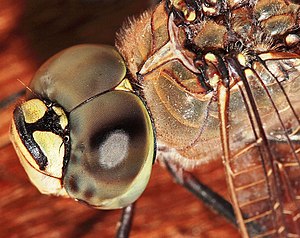
The compound eye of a dragonfly

Good fliers like flies or honey bees, or prey-catching insects like praying mantis or dragonflies, have specialized zones of ommatidia organized into a fovea area which gives acute vision. In the acute zone the eye are flattened and the facets larger. The flattening allows more ommatidia to receive light from a spot and therefore higher resolution.

There are some exceptions from the types mentioned above. Some insects have a so-called single lens compound eye, a transitional type which is something between a superposition type of the multi-lens compound eye and the single lens eye found in animals with simple eyes. Then there is the mysid shrimpDioptromysis paucispinosa. The shrimp has an eye of the refracting superposition type, in the rear behind this in each eye there is a single large facet that is three times in diameter the others in the eye and behind this is an enlarged crystalline cone. This projects an upright image on a specialized retina. The resulting eye is a mixture of a simple eye within a compound eye.

Another version is the pseudofaceted eye, as seen in Scutigera. This type of eye consists of a cluster of numerous ocelli on each side of the head, organized in a way that resembles a true compound eye.

The body of Ophiocoma wendtii, a type of brittle star, is covered with ommatidia, turning its whole skin into a compound eye. The same is true of many chitons.